About MICROFIL®

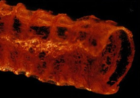
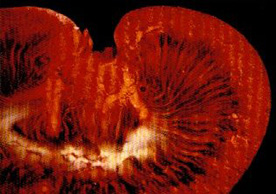
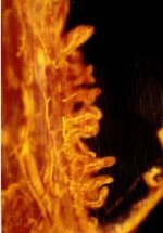
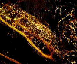
MICROFIL® compounds will fill and opacify microvascular and other spaces of non-surviving animals and post-mortem tissue under physiological injection pressure. The continuous, closed vascular system lends itself to flow through injection or perfusion techniques. Following injection, MICROFIL compounds cure to form a three-dimensional cast of the vasculature.

MICROFIL® MV-series compounds are available in five radiopaque colors, as well as clear. MV-series compounds require either an alcohol-methyl salicylate or glycerin clearing sequence, whereby the refractive index of the clearing solution is the same as the refractive index of the tissue. This allows for microscopic examination of a selected vascular bed.

Advantages offered with MICROFIL® compounds over previously available rubber injection materials include:

  • Complete filling with minimal shrinkage, to enhance vessel continuity and to produce in cleared preparations a vivid, optically cleared specimen that allows a precise study of the microcirculation.
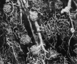
  • Color diversity, to provide delineation within the circulatory tree for microscopic examination and photographic illustration.

Areas of Investigation

In physiology, MICROFIL® visualization provides a means for establishing the precise vascular architecture of specific organs, allowing comparison between normal and abnormal structure.

In surgery, visualization of the microcirculation and microanatomy is leading to improved surgical techniques in the repair of nerves, tendons, and blood vessels.

In gastrointestinal research, MICROFIL® compounds characterize and describe changes in vascular patterns associated with several pathological conditions.

Injected specimens, when preserved in methyl salicylate or glycerin, also serve as a definitive teaching adjunct.

MV-Series Mixing Procedure

To achieve a viscosity level suitable for injection of the microcirculation, it is necessary to blend the MV compound with an equal quantity (by weight) of MV-Diluent. Volume mixing requires 5ml of diluent for every 4ml of compound. The mixture of compound and diluent is catalyzed with 5% (by weight or volume) of MV Curing Agent. Viscosity ranges from 20 to 30 centipoise. Working time is 20 minutes and begins with the addition of curing agent.

Physical Properties of MICROFIL® Compound

  MV-112
MV-117
MV-120
MV-122
MV-130
MV-132 MV-Diluent
Color White Orange Blue Yellow Red Clear Clear
Specific gravity 1.04 1.02 1.00 1.04 1.02 1.00 0.92
Viscosity, centipoise 35 25 25 25 30 20 5
Gel time, minutes 90 90 90 90 90 90 -
Useful shelf life, months 4 4 4 4 4 4 Indefinite

Notes

  1. Viscosity measured with a Brookfield Model LVF Viscometer, with a No. 2 spindle at 30RPM.
  2. Gel time measured on a blend (by weight) of one part MV compound and one part MV-Diluent followed by addition of 5% MV Curing Agent. Gel time is time required for mixture to cease flowing.
  3. Shelf life is in excess of four months. For materials held beyond four months, it is possible to run the following static test to determine acceptability:
    1. Mix 5 grams of MV compound with 5 grams of MV-Diluent in a vial.
    2. Add 10 drops (medicine dropper) of MV Curing Agent.
    3. Cap, shake, and refrigerate overnight.
    4. The mixture should gel in the vial after this procedure to assure a cure in subsequent animal injections.

Catalyzed mixtures will form an elastomeric gel after 90 minutes at room temperature. Curing takes place with non-exothermic cross-linking and minimal volume change.

It has been possible to refrigerate specimens immediately after injection and still obtain complete cure after overnight aging. The procedure decreases odor level for subsequent sectioning.

Perfusion Techniques

Two techniques of tissue clearing are described below. Alcohol-methyl salicylate clearing produces a stiffer tissue, which, from an aesthetic point, provides a pleasing view for gross observation. Glycerin clearing produces a more flexible tissue, allowing easier manipulation for a given vessel.

Alcohol-methyl salicylate clearing

Non-wetting features of MICROFIL® compounds prevent any interaction with blood. Therefore, in the non-surviving animal, a selected vascular bed can be readily perfused without prior washout of blood. Heparinization to maintain blood fluidity, however, has been used to realize improved injection preparations.

For the injection of blood vessels from vascular beds removed post-mortem, washout of clotted blood with saline is advisable.

Selected vascular beds are perfused through their accessible artery and drained through a similar vein. Infusion pressures will vary with the animal’s mean systemic pressure. For organs from the dog, cat, rat, and man, a pressure of 150mm.Hg for arterial filling has been used, and 25 to 50mm.Hg for venous fillings.

MICROFIL® injetion of a rat trachea

MICROFIL MV-130
(Red) injection of rat trachea.
Courtesy Robert A. Acland, University of Louisville

MICROFIL® injetion of a rat kidney

MICROFIL MV-130
(Red) injection of rat kidney.
Courtesy Robert A. Acland, University of Louisville

Alcohol-methyl Salicylate Clearing Sequence

First Day Immerse in a 25% solution of ethyl alcohol.
Second Day Immerse in a fresh solution of 50% ethyl alcohol.
Third Day Immerse in a fresh solution of 75% ethyl alcohol.
Fourth Day Immerse in a fresh solution of 95% ethyl alcohol.
Fifth Day Immerse in absolute ethyl alcohol.
Sixth Day Immerse for 12 to 24 hours in methyl salicylate.

If tissue has not cleared, return to 95% ethyl alcohol stage and repeat final steps of clearing procedure.

Notes

  1. At the 50% ethyl alcohol concentration, tissue specimens may be bleached with 6% hydrogen peroxide for one day. After bleaching, continue the normal clearing procedure. Peroxide bleaching permits greater depth perception; however, this procedure must be considered against some loss in color contrast.
  2. In the final stages of ethyl alcohol-methyl salicylate, the clearing liquid may have a cloudy appearance. Addition of a small amount of 10% ethyl alcohol will alleviate this condition.

After the vascular bed is perfused and the MICROFIL® injection mass allowed to cure overnight at room temperature, the tissue is subjected to the clearing sequence described in Table 2. Thin tissues may be cleared without sectioning, but thicker organs, such as kidney or brain, should be cut into 1-centimeter slices before immersion. The alcohol-methyl salicylate clearing technique is applicable to all types of tissue with the exception of brain tissue. In the case of brain tissues, it is necessary to allow two days for each step, with an alcohol solution change every day.

Glycerine Clearing

The animal is anesthetized with Nembutal, 25 mg/Kg i.v., at the same time is is heparinized, to ensure effective removal of its blood volume during perfusion.

A midline incision exposes the abdominal viscera from the sternal notch to symphysis. Following the placement of an abdominal retractor, the thoracic cage is opened rapidly, the thoracic aorta isolated, and a polyethylene cannula inserted distally. The arterial cannula is connected to a sine wave perfusion pump and, prior to instigating perfusion, the right atrium is opened to serve as a drain vent.

The animal is perfused with saline until all of the visceral blood volume is flushed out and the perfusate drained through the arterial vent is essentially free of blood. Adequate perfusion is characterized by severe blanching of all visceral organs.

During perfusion, the curing agent is added to the MICROFIL® injection mass. When perfusion is complete, the silicone rubber (e.g., MV-130 Red) is infused through the aortic cannula by syringe. When filling is complete, all organs have a rich, red coloration. MICROFIL® compound infusion is continued until the injection mass flows freely from the atrial vent. The atrium and arterial cannula are then clamped and the animal is placed under refrigeration at 4 degrees C overnight, to allow polymerization.

On the following day, specimens are taken by careful dissection, and placed in a 50% mixture of water and glycerin. At successive 24-hour intervals, the glycerin concentration is raised to 75%, then 85%, and finally pure glycerin. This procedure clears the tissue so that microscopic examination readily allows three-dimensional visualization of the vascular bed.

Monkey jenjunum perfused with MICROFIL®

Cross-sectional view of monkey jenjunum perfused with MICROFIL MV-118 (Maroon)* followed by MV-122 (Yellow).

Courtesy D. G. Reynolds, Walter Reed Army Institute of Research

* MV-118 is no longer available and has been replaced by MV-130 (Red).

Additional Notes

  1. All MV-series compounds are compatible with one another. Therefore, it is possible to mix colors together to suit your needs (e.g., mix MV-120 Blue with MV-122 Yellow to produce a green compound).
  2. Faster cure rates are possible by replacing the conventional MV Curing Agent with 2% ethyl silicate and 1% stannous octoate. Using this cross-link and curing agent combination decreases working time to 5 minutes with complete cure in 20 minutes. If you desire this type of cure system, please specify when placing your order. There is no extra charge for substituting this cure system in place of MV Curing Agent.
  3. Occasionally it may be necessary to decrease viscosity by changing the mix ratio to either 2 or 3 parts MV-Diluent for each part MV compound. If your study requires such action, the correct level of MV Curing Agent is 10% (by weight) of the amount of MV compound used.
  4. Store MICROFIL® kits at room temperature for maximum retention of pigment dispersion. Keep all containers tightly capped. If pigment settling occurs, it is better to lightly shake the MICROFIL® compound container and decant this portion. Stirring the container may put an agglomerated pigment particle into the system, which can be detrimental to perfusion.
  5. If catalyzed material should spill on clothing, the best available solvent is MV-Diluent. To facilitate removal of cured compound, swelling and softening will occur on contact with an aromatic solvent such as toluene or xylene, or chlorinated solvents such as trichloroethylene.
Human coronary artery filled with MICROFIL®

Vasa vasorum of the human coronary artery filled with MICROFIL compound.

Courtesy of A. C. Barger, R. Beeuwkes III, L. L. Lainey and K. J. Silverman, Harvard Medical School

Human coronary artery filled with MICROFIL®

Arterial injection of a dog kidney using MICROFIL MV-112 (White). Demonstrates filling of glomeruli and peritubular capillaries at 50x magnification.

Courtesy A. C. Barger, Harvard Medical School

While the information herein is believed to be reliable, Flow-Tek does not guarantee its accuracy. Users are urged to perform their own tests. MICROFIL® products are sold without warranty, and various patents may be pertinent to their use and to the use of compositions containing them. The information contained herein is not intended as a recommendation to use MICROFIL® products so as to infringe on any patent. Flow-Tek assumes no liability for the user’s violation of patent or other rights.